Imaging Residual Inflammatory Cardiovascular Risk | Antoniades et al
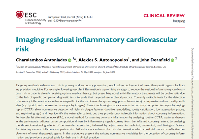
Targeting residual cardiovascular risk in primary and secondary prevention, would allow deployment of novel therapeutic agents, facilitating precision medicine. For example, lowering vascular inflammation is a promising strategy to reduce the residual inflammatory cardiovascular risk in patients already receiving optimal medical therapy, but prescribing novel anti-inflammatory treatments will be problematic due to the lack of specific companion diagnostic tests, to guide their targeted use in clinical practice. Currently available tests for the detection of coronary inflammation are either non-specific for the cardiovascular system (e.g. plasma biomarkers) or expensive and not readily available (e.g. hybrid positron emission tomography imaging).
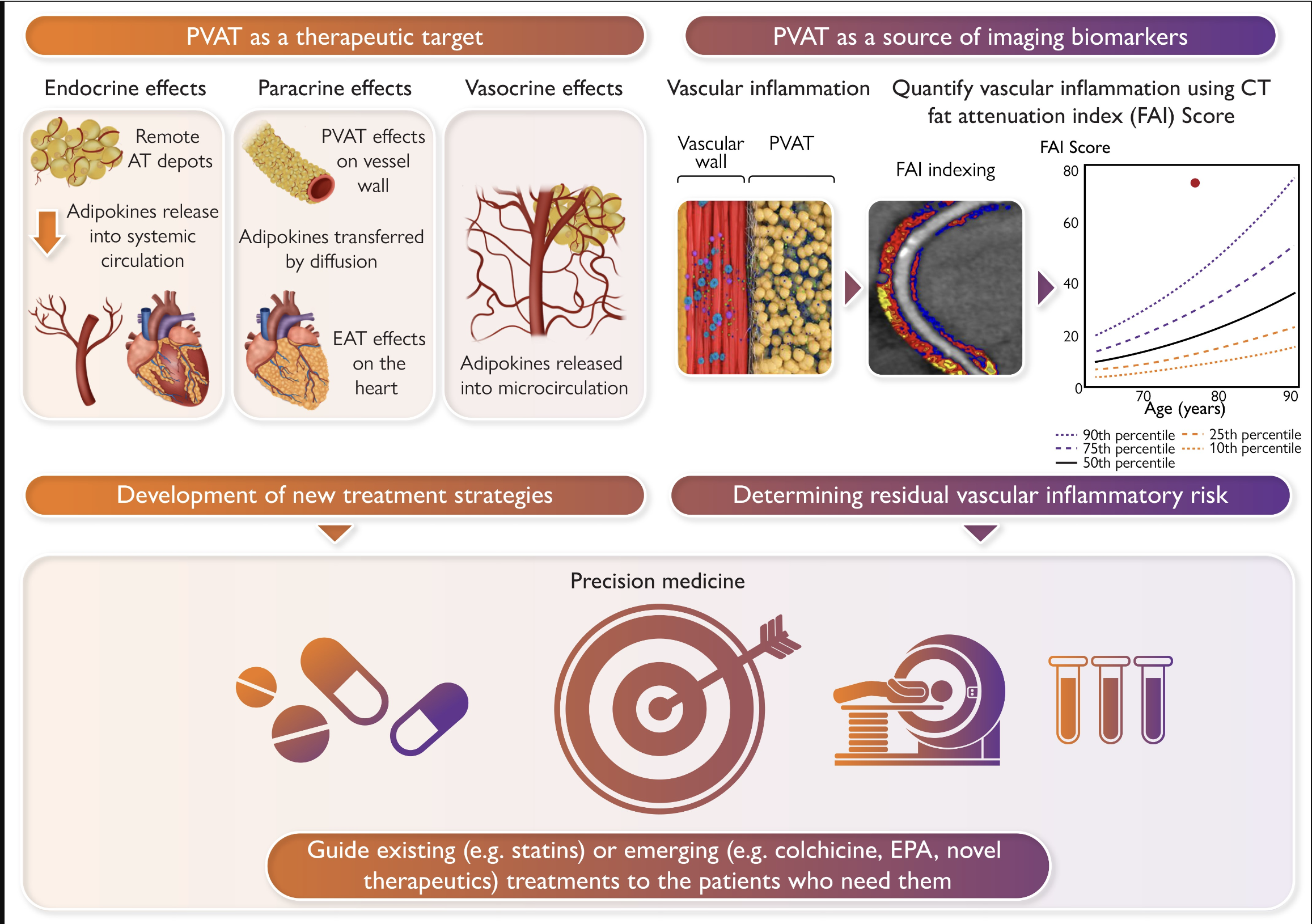
Recent technological advancements in coronary computed tomography angiography (CCTA) allow non-invasive detection of high-risk plaque features (positive remodelling, spotty calcification, low attenuation plaque, and napkin-ring sign) and help identify the vulnerable patient, but they provide only indirectly information about coronary inflammation. Perivascular fat attenuation index (FAI), a novel method for assessing coronary inflammation by analysing routine CCTA, captures changes in the perivascular adipose tissue composition driven by inflammatory signals coming from the inflamed coronary artery, by analysing the three-dimensional gradients of perivascular attenuation, followed by adjustments for technical, anatomical, and biological factors. By detecting vascular inflammation, perivascular FAI enhances cardiovascular risk discrimination which could aid more cost-effective deployment of novel therapeutic agents. In this article, we present the existing non-invasive modalities for the detection of coronary inflammation and provide a practical guide for their use in clinical practice.