Caristo Symposium at SCCT 2023 – Raising the bar for coronary artery disease management

The Caristo team were delighted to meet with the CCTA community at the Society of Cardiovascular Computed Tomography (SCCT) Annual Scientific Meeting, which was held in Boston from July 27-30.
AI/Machine Learning Workshop
Caristo was invited to take part in the AI/Machine Learning workshop. Bringing together clinical, technical and industry experts, the workshop gave delegates a unique insight into world of medical imaging software development, particularly focusing on the key clinical, data science, regulatory and commercial decisions that must be made. Delegates also heard about the latest advances in CCTA and the role that AI is playing in both hardware and software development. Dr Cheerag Shirodaria, Co-founder & Chief Development Officer, Caristo, gave a presentation entitled “coronary inflammation, the final frontier for cardiovascular risk prediction”, and took part in a panel discussion with other leading Industry experts. Interest in the advances being made in software development and the role and AI is playing was palpable throughout the workshop, as was the enthusiasm over the role that quantitative coronary inflammation assessment is playing in advancing the personalisation of clinical decision making.
Caristo Sponsored Symposium
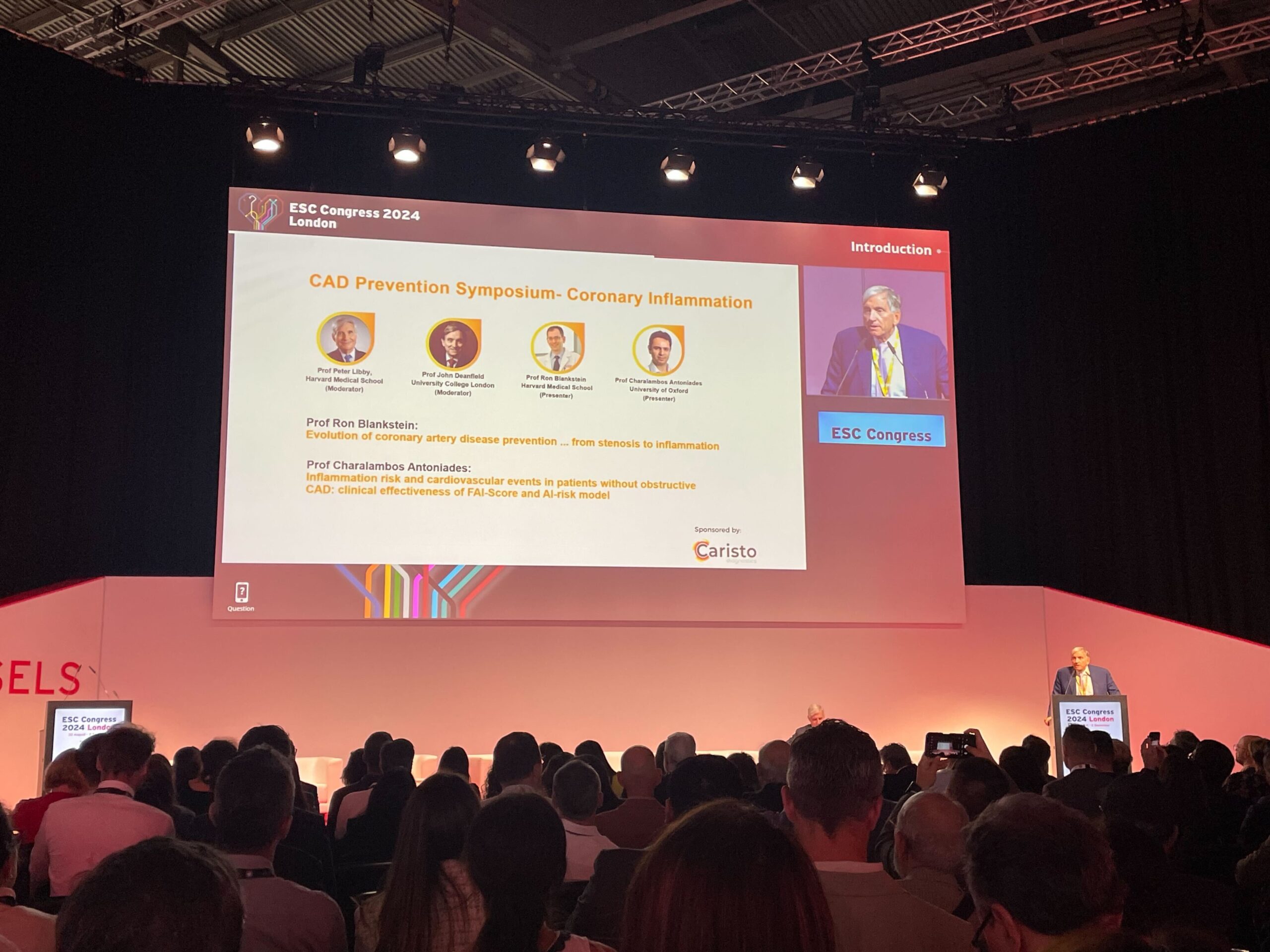
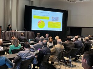
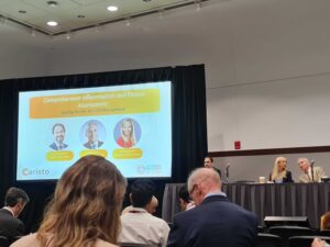
SCCT gave us the platform and opportunity to extend the conversation on coronary inflammation and plaque through a sponsored symposium on day 2 of the conference. We had a renowned panel of speakers:
- Prof Peter Libby, from Harvard Medical School, moderated the panel and opened the discussion by presenting the role of inflammation in initiating atherosclerosis and the biological mechanisms whereby adipose tissue is an important indicator of coronary risk.
- Prof Ronak Rajani from King’s College London, spoke about the science behind our CaRi-Heart Technology that reports the Fat Attenuation Index (FAI) score and plaque characteristics. As a clinical user of the CaRi-Heart technology in the UK, he also discussed the impact on the patient’s treatment pathway and results from the recent NHS AI Award.
- Dr Brittany Weber from Brigham and Women’s Hospital Boston presented interesting clinical research cases on the use of FAI for CAD diagnosis, with a special focus on its application in cardio-rheumatology.

Watch the complete symposium:
Discussions on new CT imaging techniques and coronary inflammation:
Prof. Charalambos Antoniades, from the University of Oxford and also Co-Founder and Chief Scientific Officer for Caristo, was invited by the organisers to give two presentations. In a session entitled “AI and Machine Learning: Coronary Imaging”, his talk was entitled “AI/Health and Innovation”. Here, he explained that AI is the ability of a computer to think and learn, that AI is now present in many aspects of our lives, and AI-assisted cardiac imaging will become the new norm in clinical practice, as, if deployed correctly, it will be faster and better. Requirements for its establishment as an accepted clinical tool are large-scale imaging datasets with reliable outcome collection to train the AI models, on the basis of which strictly quality-controlled, regulatory-cleared tools can then be developed.
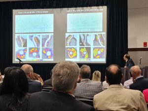
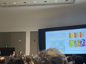
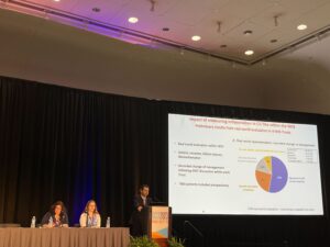
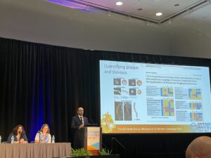
In a session entitled “Technical advances in coronary atherosclerosis imaging”, his talk was on “Fat and inflammation imaging”, Prof. Antoniades explained that new CT imaging techniques, based on assessing the attenuation of pericoronary fat, are the only method suitable for routine clinical practice which can non-invasively detect coronary artery inflammation. He reviewed the evidence for this, which is now based on many 1000s of patients and hundreds of published manuscripts, including a meta-analysis, all showing that this method provides powerful indices of coronary risk. He emphasised the importance of standardisation, which takes into account many different patient- and scanner-related parameters. As a result of this, raw measurements of fat attenuation become much more powerful, in the form of the Fat Attenuation Index (FAI) -Score and CaRi-Heart Risk, which are fully validated, regulatory approved tools that can bring the benefits of this new technology to all patients undergoing cardiac CT scans. He also explained that such coronary inflammation measurements provide risk estimates that are mostly independent from information derived from coronary plaque assessment. Finally, he reported preliminary results from a real-world assessment in the NHS, where deployment of the CaRi-Heart risk assessment tool led to a change in clinical management in 45% of the 800 patients included.
The importance of inflammation in adipose tissue, that is detected by quantified by Caristo’s CaRi-Heart technology, was also highlighted in many other talks during the meeting, including podium presentations by key opinion leaders in cardiac CT imaging, and in the presentations selected for the Young Investigator Award.