Detecting Human Coronary Inflammation by Imaging Perivascular Fat | Antonopoulos AS, Antoniades C. et al
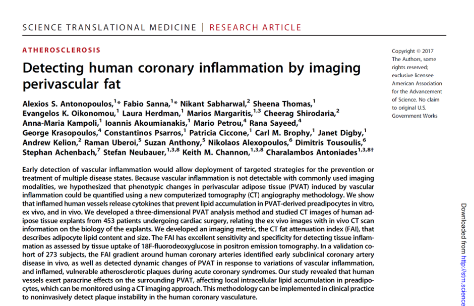
To determine risk of future coronary artery disease, calcium content in vascular plaques is typically evaluated by coronary calcium scoring, which uses computerized tomography (CT) imaging. To detect inflammation and subclinical coronary artery disease (soft, noncalcified plaques), Antonopoulos et al. developed an alternative metric called the perivascular CT fat attenuation index (FAI). The perivascular FAI uses CT imaging of adipose tissue surrounding the coronary arteries to assess adipocyte size and lipid content. Larger, more mature adipocytes exhibit greater lipid accumulation, which is inversely associated with the FAI.
Inflammation reduces lipid accumulation and slows preadipocyte differentiation. Imaging pericoronary fat in human patients after myocardial infarction revealed that unstable plaques had larger perivascular FAIs than stable plaques and that the FAI was greatest directly adjacent to the inflamed coronary artery. The perivascular FAI may be a useful, noninvasive method for monitoring vascular inflammation and the development of coronary artery disease.