Reimagining Cardiovascular Imaging in ASCVD Trials: How Biomarkers Are Redefining Risk and Response

Clinical trials for atherosclerotic cardiovascular disease (ASCVD) are increasingly turning to advanced imaging to uncover what conventional tools might miss — particularly perivascular inflammation. While traditional scoring systems like calcium burden offer structural insights, they often fail to detect early biological activity that could predict future cardiovascular events.
According to a report, the ASCVD market reached $24 billion across the US, EU4, UK and Japan in 2024 and is projected to grow to $32.3 billion by 2035 — an expansion fueled by the rising use of statins, antiplatelet therapies and minimally invasive interventions, as well as growing interest in gene and regenerative therapies.
Yet as the disease becomes better understood, so too must trial strategies evolve.
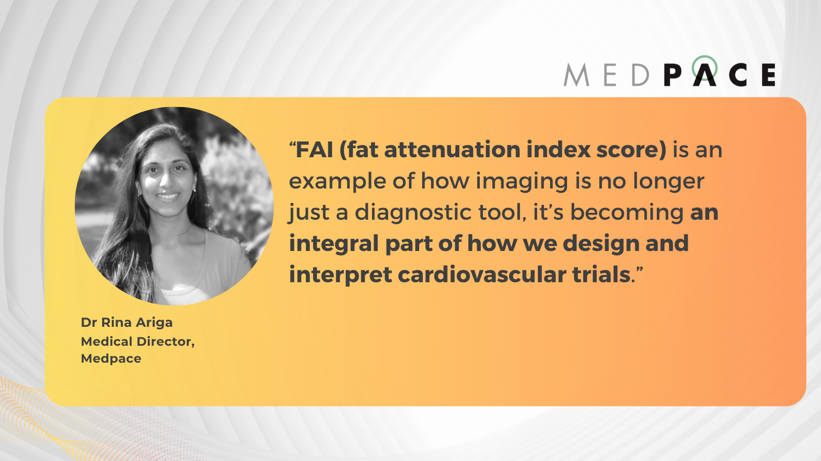
In a recent webinar, Dr. Charalambos Antoniades, Co-Founder and Chief Scientific Officer of Caristo Diagnostics, joined Medpace experts Dr. Rina Ariga, Medical Director, and Dr. Andrea Flannery, Director of Clinical Trial Management at Medpace, to discuss how imaging biomarkers like the fat attenuation index (FAI) and emerging techniques such as radiotranscriptomics are changing how ASCVD trials are designed and interpreted.
Read on to learn how this expert panel is advancing imaging strategies that improve risk prediction, clarify biological mechanisms and support operational planning in cardiovascular trials.
Rethinking Inflammation Detection in Cardiovascular Risk
Dr. Antoniades opened his talk with a fundamental question in cardiovascular prevention: should we pursue a cost-effective, one-size-fits-all approach like the poly-pill, or invest in precision medicine that targets the right treatments to the right patients?
While current practice still relies heavily on risk-factor-based calculators, Dr. Antoniades argued that meaningful implementation of precision medicine requires integration of genetics, plasma biomarkers and imaging biomarkers.
He explained that coronary artery disease affects about 6.5% of the population, typically identified when patients develop symptoms. However, nearly 60% of heart attacks occur in individuals without symptoms or detectable obstructive disease. This underscores the need to detect disease earlier, even before significant plaque formation, by identifying inflamed arteries and vulnerable plaques.
Coronary CT angiography (CCTA) now serves as the first-line test in many cases of chest pain. Findings from the ORFAN study — a prospective study of 40,000 patients followed for three years after undergoing CCTA — showed that only 18% had obstructive disease, yet two-thirds of heart attacks occurred in patients without obstruction. That insight changed the thinking around inflammation as a therapeutic target and a diagnostic gap.
He cited several clinical trials — CANTOS, LoDoCo2 and COLCOT — which confirmed that targeting inflammation reduces cardiovascular events, leading to colchicine’s FDA approval for cardiovascular risk reduction in 2023.
Still, a key challenge remains: identifying the subset of patients with inflamed coronary arteries. Plasma biomarkers reflect systemic inflammation, not localized arterial inflammation.
To bridge that gap, Dr. Antoniades and his team at the University of Oxford studied how inflamed arteries alter the surrounding perivascular adipose tissue (PVAT). When arteries are inflamed, PVAT cells shrink and lose lipid content — a dynamic change visible even before plaque forms (Figure 1).

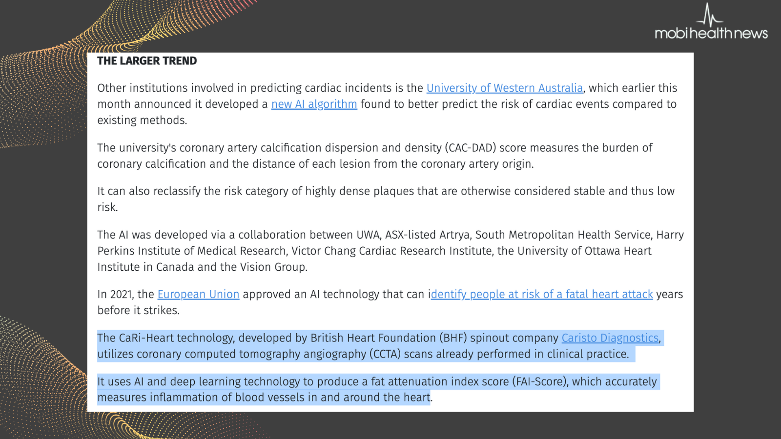
To make this insight clinically actionable, his group developed FAI — a CT-based “virtual biopsy” that reveals inflammation by analyzing the attenuation patterns of PVAT.
Normal, non-inflamed arteries appear yellow, whereas inflamed regions appear red. In some cases, patches of blue indicate fibrosis and edema linked to specific inflammatory pathways.
Dr. Antoniades spoke about a photon-counting CT research facility (Figure 2) integrated with a cardiac catheterization lab (cath lab) to validate FAI and support its clinical utility compared to intravascular imaging.

The team refined the method further, developing an FAI score that adjusts for anatomical and technical variables, allowing broad application across coronary vessels. This score has been regulator-approved as a biomarker of coronary inflammation in Europe, the UK and Australia, and was incorporated into a European Society of Cardiology (ESC) clinical consensus statement on coronary inflammation imaging.
Validating FAI: What the Data Shows
Dr. Antoniades outlined how large-scale studies have established FAI’s value as a cardiovascular imaging biomarker.
In the CRISP-CT study of 4,000 patients in the US and Germany, FAI improved risk prediction beyond traditional markers like calcium scores and plaque burden. It increased the C-statistic — a measure of a model’s ability to differentiate between patients with and without events — by 5.4 to 7.5 points.
To assess generalizability, his team launched the ORFAN study, aiming to enroll 250,000 patients undergoing coronary CT. As of mid-2025, over 210,000 patients have been recruited.
Early findings from the UK arm of the ORFAN study revealed that patients with FAI scores above the 75th percentile were at markedly higher risk for adverse outcomes. Specifically, those without obstructive coronary disease faced a tenfold greater risk of cardiac death, while those with obstructive disease had a fivefold increase. Inflammation scores also correlated with a 3.5 to 5.3 times higher likelihood of non-fatal myocardial infarction across all three coronary arteries. Additionally, high FAI values were linked to a greater incidence of ischemic heart failure — even in patients without prior myocardial infarction.
These outcomes were observed even in patients with no visible plaque or a calcium score of zero. Data from ORFAN further reported hazard ratios exceeding 10 for cardiac death in patients with minor or no plaque but high FAI, reinforcing the biomarker’s independent prognostic value.
Dr. Antoniades noted that FAI also helps monitor treatment effects. In patients with psoriasis, biologic therapies such as anti-TNF and anti-IL agents have led to notable reductions in FAI. – Post-STEMI (ST-elevation myocardial infarction) patients — those recovering from a severe heart attack caused by a fully blocked artery — showed significant FAI decreases within six months. However, 20% remained non-responders, a group that could likely benefit from novel therapeutics.
Statin therapy has shown a 30% to 40% reduction in FAI within one year, sustained for at least three years. In a 12-week Phase II randomized clinical trial, orticumab, a monoclonal antibody targeting oxidized LDL, significantly lowered FAI in all three coronary arteries — a promising indicator for Phase II trials.
Dr. Antoniades also pointed to an observational study in breast cancer patients receiving radiotherapy. While calcium scores rose — typically considered a negative signal — inflammation levels dropped across all coronary arteries. He suggested this may reflect plaque stabilization after tumor removal rather than elevated cardiovascular risk.
Collectively, these findings point to FAI as a promising early marker that could add value alongside conventional imaging and clinical evaluations, particularly in identifying patients who may not be flagged through standard measures.
Toward Smarter Trials: Radiotranscriptomics and AI Integration
To refine patient selection for cardiovascular trials, Dr. Antoniades emphasized the need to help personalize cardiovascular risk prediction. Dr. Antoniades introduced CaRi-HEART, an AI-powered platform that combines CCTA with clinical and biological data to estimate absolute cardiac risk.
Trained on cohorts in the US and Germany and validated in the UK, CaRi-HEART achieved a C-index of 0.84 for predicting cardiac mortality. In the UK’s National Health Service’s (NHS) use, the tool influenced treatment decisions in 45% of patients — prompting statin initiation in 24%, dose increases in 13% and referrals in 8%. These shifts underscore its potential to guide trial enrichment and target patients most likely to benefit from intervention.
To push this further, Dr. Antoniades’ team developed the Heart, Vessels and Fat (HPF) Library — a radiotranscriptomic dataset linking visible fat changes on CT to inflammatory signals identified via RNA sequencing. This work aims to connect imaging features with underlying biology, supporting more precise patient selection and early efficacy insights in clinical trials.
Read the full story to on Xtalks.com to learn how FAI-Score is advancing the design and interpretation of future ASCVD trials
###


